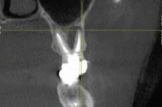
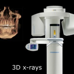
This level of detail makes it possible to evaluate the jawbone, tooth roots, and jaw joints (TMJ) with exceptional accuracy—supporting safer implant placement, improved diagnosis, and more predictable treatment outcomes.
3D X-Rays and Guided Implant Planning
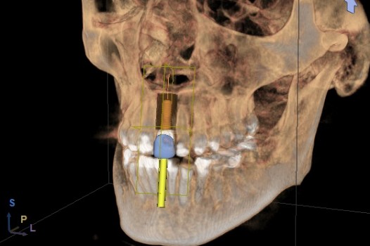
With 3D CBCT X-ray imaging, Dr. Klim can capture highly detailed, three-dimensional views of the teeth, jawbone, nerves, and surrounding structures. Using this data, advanced software allows Dr. Klim to virtually plan dental implant placement with exceptional precision.
Once the digital planning phase is complete, the information is sent to a dental laboratory or an in-office milling system. From this plan, a custom surgical guide (also called a stent) is fabricated specifically for the patient.
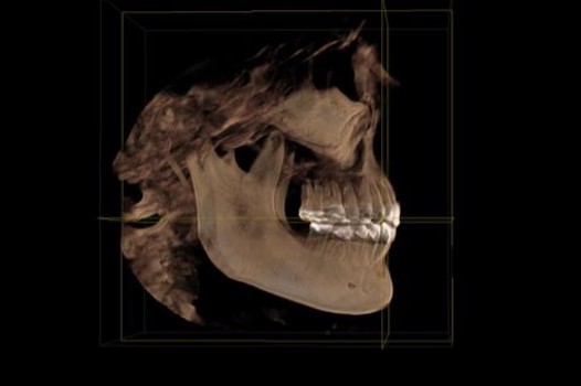
During the procedure, the guide is placed securely in the patient’s mouth. The guide’s precise opening directs the instruments, allowing the bone to be prepared and the implant to be placed in the exact planned position, improving accuracy, safety, and long-term results.
Other Uses of 3D CBCT X-Rays
Dental implant planning is just one of the many benefits of 3D CBCT imaging.
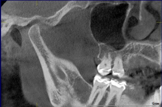
Because a 3D X-ray provides significantly more information than traditional 2D X-rays, Dr. Klim can also use this technology to:
-
Diagnose root fractures and internal tooth damage
-
Detect infections or pathology that may not be visible on standard X-rays
-
Evaluate the jaw joints (TMJ) for TMJ/TMD diagnosis and treatment
-
Assess anatomical factors related to sleep apnea
-
Support advanced treatment planning for complex dental conditions
By viewing the jaw, joints, and surrounding structures in full detail, 3D CBCT X-rays help ensure more accurate diagnoses and more predictable treatment outcomes.